Healthy eating with diabetes
Read more information on the Diabetes UK website
10 tips for healthy eating with Diabetes
Gestational diabetes (GDM) is high blood sugar (glucose) developed during pregnancy and usually disappears after giving birth (post-delivery). It can happen at any stage of pregnancy but is more common in the second or third trimester. It happens when your body cannot produce enough insulin – a hormone that helps control blood sugar levels – to meet your extra needs in pregnancy.
Key features of GDM:
Any woman can develop gestational diabetes during pregnancy, but you have a higher risk if:
Some women with no risk factors at all will also develop GDM. The key message is that GDM can happen through no fault of your own but there are lots of things you can do to help manage it.
Most women with GDM have otherwise normal pregnancies with healthy babies. However, gestational diabetes can cause problems such as:
If you have gestational diabetes, the risks associated with GDM can be reduced by controlling your blood sugar levels.
You’ll be given a blood sugar testing kit and asked to test a minimum of four times a day. The instructions are included and you will be sent links to the Agamatrix user video.
Blood glucose targets:
Gestational diabetes normally goes away after birth. But woman who have had it are more likely to develop:
You should have a blood test to check for diabetes 13 weeks after giving birth, and once every year after that if the result is normal.
See your GP if you develop symptoms of high blood sugar, such as increased thirst, needing to wee more often than usual, and a dry mouth, losing weight, repeated thrush, slow healing wounds– do not wait until your next test.
You should have the tests even if you feel well, as many people with diabetes do not have symptoms.
There is a type 2 prevention programme which your GP can refer you to or you can self refer to.
A healthy diet is an important part of a healthy lifestyle at any time but especially for women who are pregnant or planning a pregnancy. What you eat and drink plays a key role in managing your blood glucose levels during pregnancy when you have GDM. The right balance of a variety of food is also important to make sure you and your developing baby get the nutrition you both need.
We base our healthy eating advice around the Eatwell guide, which you can find links to below. The Eatwell Guide divides the foods and drinks we consume into five main groups. Cultural and ethnic versions of the Eatwell Guide have been created which include foods commonly eaten in African and South Asian communities. Try to choose different foods from each of the food groups to help you get a wide range of nutrients that your body needs to stay healthy and work properly.
After monitoring glucose levels, the second step for managing gestational diabetes involves looking at your diet and exercise and making changes where possible to help control your blood glucose levels.
We recommend you base your meals around the portion plate model which is pictured below. This includes half a plate or vegetables or salad, a quarter of a plate of starchy carbohydrate foods, and a quarter of a plate of protein.
Counting the amount of carbohydrates in your meals is not usually necessary, however if you find it helps, here is a guide, per meals:
Note: This is just a guide, everyone has different degrees of insulin resistance with their GDM. By regular blood glucose monitoring you’ll be able to adjust your diet as you need to.
Carbohydrates have the biggest impact on our blood glucose levels. Your body breaks down carbohydrates into glucose (sugar).
It is important to not cut these foods out completely as they are the main source of energy for your body and your growing baby. They also provide important vitamins and can be a source of fibre.
You can eat all types of carbohydrate, but some will raise your blood glucose levels more than others, even when an appropriate portion size is eaten. Try to eat more of the higher fibre/low GI carbohydrates, as per the table below.
Our starchy carbohydrates are foods within the yellow section of the Eatwell guide. Examples include:
Many women find that breakfast cereals cause blood glucose levels to rise too much and must avoid them completely during pregnancy.
Try a lower carbohydrate breakfast such as natural or Greek yoghurt with one portion of fruit or eggs/avocado/cottage cheese, with one slice of seeded bread.
All food that contains carbohydrate have a glycaemic index value (GI). This is the speed that the body breaks down carbohydrate into glucose and absorbs it into the blood.
Low GI foods are the best choices as they are absorbed and raise the blood glucose more slowly so can help to keep your blood glucose levels within the target range and regulate your appetite much better. High GI foods are absorbed quicker and raise the blood glucose quickly.
Therefore we recommend that you try and choose foods from the Low GI food list.
The table below shows you some examples of low and high GI foods.
| Refined carbohydrates to avoid (High glycaemic index (GI)) | Try instead (Low glycaemic index (GI)) |
|---|---|
All white breads:
|
|
Low fibre and sugar coated breakfast cereals:
|
|
Rice, pasta, grains:
|
|
Processed potato products:
|
|
Fats are needed in the diet as they play an important role with different aspects of health. Such as providing energy, warmth, protection of the internal body and key fat-soluble vitamins. However, it is important to note that all fats are very high in calories which can contribute to unhelpful weight gain.
Examples of food sources that are high in fats include:
We generally don’t need to deliberately add high fat foods into our diet as most people get enough fats within their normal diets. However, it is appropriate to try to keep our intake of high fat foods to 2-4 portions per day. Examples include a tablespoon of oil or butter, half an avocado, or 20g of nuts.
All fats are high in calories, but not all fats are the same in other ways, such as their impact on cholesterol level. It is important for everyone to try to limit their intake of saturated fats, such as those from animal products, and swap to unsaturated fat where possible, such as those in nuts, plant based oils and avocado.
Free sugars are sugars added to food such as in cakes, biscuits, chocolate and desserts. They are also naturally in honey, syrups and unsweetened fruit juices. However, the term free sugars excludes lactose in milk and milk products, and the sugars in whole fruit.
All sugars are a form of carbohydrate and therefore provide a rapid source of energy for our body, but foods high in free sugars often provide very little other nutritional benefit.
Free sugars also have a very quick impact on our blood glucose levels. This is normally unhelpful and pushes the levels above the target ranges unless our blood glucose levels are too low to start . It is important to reduce your intake of free sugars as much as possible, including in drinks.
For more information on this please check: Exercise in pregnancy - NHS
Weight gain is generally inevitable during pregnancy due to all the physical changes that happen to help your baby grow and develop, but it is important to try not to gain too much weight throughout your pregnancy, particularly if you were overweight before.
There are certain foods that are best to avoid while you are pregnant as they can put your baby’s health at risk. These include some types of cheese and raw or uncooked meat. For more details, please see websites below.
Try to eat regular meals (breakfast, lunch, and evening meal). Spread your intake over the day to help to manage your blood glucose levels.
If you skip meals, you might be more likely to overeat at the next meal or end up on snacking on things that can have a big impact on your blood glucose.
Avoid eating carbohydrates late in the evening. Having your dinner too late at night can affect your fasting blood glucose the next day. If you can, try to have your dinner 2-3 hours before you go to bed. If you need to snack after dinner, opt for low carb options like nuts, cheese or a small portion of Greek yogurt.
© North Bristol NHS Trust. This edition published September 2025. Review due September 2028. NBT003601.
See the impact we make across our hospitals and how you can be a part of it.
Living with a long term health condition can have an impact on your emotional wellbeing. Looking after your emotional health is just as important as looking after your physical health.
Evidence* suggests there are five steps we can all take to improve our mental wellbeing. If you give them a try, you may feel happier, more positive, and able to get the most from life.
Psychological therapies, sometimes called talking therapies, can help with common difficulties like stress, anxiety and low mood. If you are registered with a GP you can refer yourself directly to free services (or you can ask your GP to refer you).
These services are also known as Improving Access to Psychological Therapies (IAPT) services and provide psychological support in a variety of ways including face-to-face, online, over the phone, and courses.
VitaMinds
0333 200 1893
refer.BNSSG@nhs.net
01225 675150
01380 731335
awp.wilts-iapt@nhs.net
01793 836836
lift.psychology@nhs.net
If you have any concerns about your mental wellbeing or physical health you can always contact your GP. If you need urgent help please call 999 or attend A&E.
If you are feeling distressed and would like a confidential listening service, you can contact the Samaritans. They are available 24 hours a day, 7 days a week.
Telephone: 116 123
Email: jo@samaritans.org
The Kidney Peer Mentor Service aims to give short term practical and emotional support to people living with kidney disease, their families, and carers. All trained peer mentors are people who have lived with kidney disease themselves, and are allocated to you based on their experiences. If you would like to find out more please email: kidneypeermentorservice@nbt.nhs.uk
We have two clinical psychologists based within the Renal Team at Southmead to support people with a wide range of issues. Sometimes people experience emotional difficulties as a direct result of adjusting to and living with kidney problems, at other times difficulties may come up independently making it more difficult to cope with the demands of living with kidney problems and treatment. If you would like an appointment with one of the renal psychologists please speak to a member of staff who will be able to refer you.
If you or the individual you are caring for need support reading this please ask a member of staff for advice.
Clinical Psychology Service
Renal & Transplantation Directorate
Gate 10 Level 6
Southmead Hospital
Westbury on Trym
Bristol
BS10 5NB
0117 414 7696
www.nbt.nhs.uk
Peritoneal dialysis catheter insertion: Placing a Tenckhoff catheter in the abdomen
Your doctor has recommended you start dialysis treatment for kidney failure because of your blood test results and how you feel. You have chosen peritoneal dialysis.
The peritoneum is a membrane that lines the organs in the abdomen (tummy). Peritoneal dialysis (PD) filters the blood to “clean” it, when your kidneys don’t work well enough to keep you healthy.
A permanent tube called a Tenckhoff catheter is placed in the lower part of your belly. This is used to put in and drain out dialysis fluid. It is a soft, flexible tube, that lies flat against your body. There is a cap on the end of the tube so no opening.
For Peritoneal Dialysis the fluid is left in your abdomen to do the dialysis for several hours, then drained out. This fluid takes away some of the waste products and excess water that are normally removed by the kidneys. Fresh fluid is then put into the abdomen.
This exchange of fluid takes about 30 minutes and you will do it 2 - 4 times a day. The fluid comes in a range of volumes and “strengths”. The type of fluid you use depends on your needs.
The catheter is inserted by a surgeon at Southmead Hospital. The operation will usually be a day case (you will not stay overnight) using general or local anaesthetic.
There are a few things to think about when choosing the position of your catheter. You will discuss this with your surgeon before the operation and the community dialysis nurse will mark the place you agree on your skin.
Before the procedure we will take swabs of your nose and groin. This is to check if you are carrying any bacteria on your skin that could cause infection of the catheter.
If the swabs are positive you may need treatment to clear the bacteria before the procedure.
The usual treatment is:
If for any reason you haven’t had the swab tests done before the procedure, they will be done when you are admitted. If treatment is required it will be started then for at least 5 days.
If you take warfarin, clopidogrel or any other blood-thinning tablets, you will need to stop taking these before the procedure. Your doctor or pre-operative assessment clinic (POAC) nurse will tell you when to stop taking them.
You will be given a date to attend the pre-operative assessment clinic (POAC). Some blood samples and swabs will be taken, and measurements of your blood pressure, pulse, temperature, and oxygen level.
Blood samples and swabs will be taken at the initial visit to your home. Before your surgery a member of the Renal Community Team will come to your house to go through the details of peritoneal dialysis (PD) including:
You will be given a date for your surgery. It is important your bowels are empty so you will be asked to take laxatives called sodium docusate and senna 3 days before the operation.
You will continue to take laxatives after the operation to make sure the PD tube won’t move because of constipation.
You will go to the medirooms.
You will go to the renal ward 8b.
Most patients are advised to take 7 days of work to recover.
Some people need more than this, particularly if you have a manual job.
There are 4 main possible complications that may happen.
Occasionally the catheter might not work. This might be because the tip of the catheter is in the wrong place or is covered by internal tissue which stops it working. At Southmead this happens in less than 7 in 100.
Bleeding can happen, mostly because of bruising around the wound. It usually stops on its own. In rare cases (less than 1 in 100) you may need surgery to stop the bleeding.
You will have intravenous (IV) antibiotics when the catheter is inserted to prevent infection. Despite this, some patients develop infection at the catheter exit site which needs further antibiotics. This happens in less than 1 in 100 cases, and very occasionally the catheter may need to be removed.
There is also a risk of infection deeper in the abdomen, causing peritonitis in the first 2 weeks after insertion. This happens in about 3 in 100 cases, and will be treated with different antibiotics.
Very rarely during the procedure the bowel is cut by mistake. This happens in less that 1 in 100 cases. It is a serious complication that may need another operation and a stay in hospital.
A community nurse will look after your ongoing care, and they will keep in contact with you. This will involve some visits to your home. A follow-up clinic appointment will be arranged with one of the renal doctors after your training.
Hospital transport can be arranged for appointments and training, but this is only for patients who meet certain criteria. We encourage you to make your own arrangements if possible.
© North Bristol NHS Trust. This edition published October 2024. Review due October 2027. NBT002131.
See the impact we make across our hospitals and how you can be a part of it.
Renal Community Team
Level 1, Gate 5
Brunel building
Southmead Hospital
BS10 5NB
Living with a long-term health condition can have an impact on your emotional wellbeing. Looking after your emotional health is just as important as your physical health.
Evidence suggests there are five steps we can all take to improve our emotional wellbeing. If you give them a try you may feel happier, more positive, and able to get the most from life.
Psychological therapies, sometimes called talking therapies, can help with common difficulties like stress, anxiety, and low mood. If you are registered with a GP you can refer yourself directly to services, or ask your GP to refer you. These services used to be known as Improving Access to Psychological Therapies (IAPT) services. They provide psychological support in a variety of ways including face-to-face, online, over the phone, and courses.
If you have any concerns about your emotional wellbeing or physical health, you can always contact your GP. If you need urgent help please call 999 or go to A&E.
If you are feeling distressed and would like a confidential listening service, you can contact the Samaritans by phoning 116 123. They are available 24 hours a day, 7 days a week.
Social prescribing is available through all GP practices. It aims to help people improve their health, emotional wellbeing, and social networking by connecting them to community activities and resources. For example, social prescribers can discuss your interests with you and help find local activities that you might want to get involved in.
The Kidney Peer Mentor Service aims to give short-term practical and emotional support to people living with kidney disease, their families, and carers. All trained peer mentors are people who have lived with kidney disease themselves, and are allocated to you based on their experiences. If you would like to find out more please email: kidneypeermentorservices@nbt.nhs.uk
We have two clinical psychologists based in the Renal Team at Southmead to support people with a wide range of issues. Sometimes people experience emotional difficulties as a direct result of adjusting to and living with kidney problems. At other times difficulties may come up independently, making it more difficult to cope with the demands of living with kidney problems and treatment.
If you would like an appointment with one of the renal psychologists, please speak to a member of staff who will be able to refer you.
© North Bristol NHS Trust. This edition published January 2025. Review due January 2028. NBT003225
See the impact we make across our hospitals and how you can be a part of it.
Possibly a Transient Ischaemic Attack (TIA): this can take place because of a temporary blockage in a blood vessel in a part of the brain.
This can be similar to a stroke with symptoms such as weakness, numbness, talking problems or trouble with your vision.
TIA is different as it only lasts between a few minutes to a few hours.
There are other possible causes for these type of symptoms and over half of the people attending the clinic turn out not to have a TIA.
About one in ten people who have a TIA develop a stroke within the next week but we can greatly reduce this with the right treatment.
The right medication can reduce your chance of a stroke, as can changes to your diet and exercise. Rarely an operation may be useful. We will discuss all this when you see us in clinic.
What may we need to do? (each case is different – you may not need all these investigations)
You can expect to be at the clinic for 3-4 hours.
The clinician who initially diagnosed you may give you medication to take. You need to take this each day until you are seen in our clinic.
There are clinics at Bristol Royal Infirmary and Weston General Hospital and weekend clinics at Southmead Hospital. If you are told to attend a TIA clinic, the TIA co-ordinator will tell you which clinic to attend.
Every day, the stroke specialist on duty will review all new referrals. If we decide that we need to see you urgently over the weekend or at a bank holiday, the co-ordinator of the TIA Service will phone you on the day tell you what time to come to the unit.
Otherwise, the TIA co-ordinator will be in touch on the next working day to arrange an appointment for you to come in. In some cases, the appointment will be a telephone consultation only. The appointment will usually be on the same day. If you have not heard from us within two working days, please ring the TIA co-ordinator on 0117 342 4800.
Please bring a list of all the prescribed tablets you take – this is very important. If there are other tablets you buy over the counter, please tell us about these. If possible, it will be helpful for the person who witnessed you having your symptoms to come to the appointment as well.
There is a Costa Coffee, a small shop and a League of Friend’s coffee shop on Level 1 of the Brunel building where you can purchase refreshments. Alternatively, you may want to bring your own provisions.
It is not safe or legal for you to drive until you have been seen in clinic. We will discuss any possible further driving restrictions related to your diagnosis with you in the clinic.
If you think you are having a stroke, don’t wait – call 999!
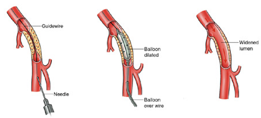
There are two types of treatment to open up blocked or narrowed arteries, without the need to make an opening in the skin. These are called “angioplasty” or “stenting,” and can be done using X-ray guidance. They are usually performed while you are awake, using local anaesthetic to numb the skin.
The first stage of your procedure is called an angiogram. This is used to make a detailed map of your arteries, because blood vessels do not show up on ordinary X-rays. A needle, followed by a thin plastic tube (catheter) is inserted into your artery to do this.
After the angiogram, a soft guidewire is inserted into the artery through the catheter. The wire must then be passed through the blockage or narrowed section of artery, if this is not possible we will discuss your options with you. A special balloon is then passed over the top of the wire. This balloon is inflated to stretch open the blockage or narrowing and allow more blood to flow through.
Once the artery has been stretched the balloon is removed and another angiogram is done to look at the result. If it is not possible to pass through the blocked artery we will discuss your options with you.
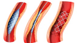
A stent is a hollow mesh tube about the size of your little finger. It is made of fine, sterile metal and is used to open an artery. When it is used, unlike an angioplasty balloon, it is left inside the artery after the procedure and remains there forever. Once it is in position the stent will not give you any discomfort or sensation. It is important to remember it is there and to tell any doctors treating you in the future that you have it. It can be placed in the artery using similar equipment to the angioplasty and usually requires no additional anaesthetic.
Stents are often used if there is a long blockage in a large artery. It may also be used if you have an angioplasty and the result is not as good as hoped for, or if the artery is not smooth enough for an angioplasty.
If with time, a narrowing that was treated with an angioplasty reoccurs, a stent can be used to decrease the likelihood of it narrowing again. Narrowing can also develop within or around a stent, or the stent could get blocked. If the stent blocks, your symptoms may return or get worse.
Most angioplasties/stents are performed by the Major Arterial Centre in Southmead Hospital. They are done by specialist radiology doctors called interventional radiologists, who are part of the multidisciplinary team involved in treating patients with vascular disease. Unless you live alone, are frail, or have certain other medical needs, you will usually go home the same day as your procedure.
The procedure is done in a specially adapted room called an “Interventional Radiology (IR) room”. Some patients will have surgery at the same time as angioplasty or stenting. This is done in a specialist operating theatre (hybrid theatre) which has X-ray equipment.
The procedure will be performed by a team of specialists. In most cases the team is led by a consultant interventional radiologist. The lead doctor may be assisted by another specialist, or a doctor training in radiology or vascular surgery. Other members of the team in the IR room will include radiology nurses and radiographers.
When the local anaesthetic is injected it will sting to start with, but this soon wears off. The skin and deeper tissues should then start to feel numb. After this the procedure should not be painful but you may feel pressure or pushing. If the procedure becomes uncomfortable we can give you some painkillers through the cannula in your arm.
As the contrast medium passes around your body you may get a warm feeling, which some people find a little unpleasant. This will soon pass and should not concern you.
Some people feel a bit of discomfort when the angioplasty balloon is inflated or when the stent is expanded. This usually passes quickly when the balloon is removed.
Every patient’s situation is different, and it is not always easy to predict how straightforward the procedure will be. For example, those with a large artery in the leg are usually straightforward and do not take long – around half an hour. Other times the vessels may be much smaller and complex, and the procedure can take 2-3 hours.
Overall there is a 1 in 100 chance an angioplasty will fail and immediately make your leg worse. In this situation you may need urgent surgery. In severely diseased arteries the risk of requiring urgent surgery is 3 in 100. If urgent surgery fails to restore blood flow to the leg you may even need an amputation.
Complications following an angioplasty are less frequent than following surgical alternatives but can include:
You will be taken back to our day case area on a trolley. Nurses in the day case area will carry out routine observations such as taking your pulse and blood pressure to make sure there are no problems. They will also look at your skin where the catheter was inserted to make sure there is no swelling or bleeding around it. In most cases you will be required to lay flat in bed for a few hours until you have recovered. If the specialist has used a closure device you will be able to sit up quite soon after your procedure.
Your specialist may make recommendations about your usual medication following the procedure. It is common to prescribe a short course (6 weeks) of tablets to help keep your angioplasty or stent open and working well whilst the artery repairs itself.
If your procedure goes as planned, most patients come into hospital, and go home from hospital, on the same day.
If you go home the same day a responsible adult should accompany you home in a car or taxi. They should stay with you at home for 24 hours. You should not drive, operate machinery, or do any potentially dangerous activities for at least 24 hours. You should wait longer if you don’t feel fully recovered.
You should not do strenuous exercise for 1-2 days.
You should check your travel insurance if you wish to travel within 4 weeks of this procedure.
The benefits you get from a successful procedure depends on many factors, especially whether you smoke, and the pattern of your arterial disease.
The results of angioplasty and stenting are most effective when they treat:
You cannot do anything to relieve the actual narrowing or blockage being treated.
You can improve your general health by doing regular exercise, stopping smoking, and reducing fat in your diet. These actions will help slow down the hardening of arteries which caused the problems in the first place, and may help you avoid the need for further treatment in the future.
Most patients will have a telephone follow-up with a clinical nurse specialist. This is a member of the specialist team.
If you have ongoing symptoms, we will arrange for you to be seen in an outpatients appointment.
If you have had a stent inserted in the thigh artery or behind the knee, the vascular team will arrange for you to have an ultrasound (arterial duplex) done at your closest hospital, around 6 weeks after your procedure. You will then be asked to attend further scans (surveillance) at regular intervals over the next 12 months. Sometimes the stent can develop narrowing without causing any symptoms. Your specialist would then discuss the pros and cons of intervention (further angioplasty).
We hope this information is helpful. If you have any questions before or after the procedure, the staff in the Imaging Department will be happy to answer them. The phone number is on your appointment letter.
The Circulation Foundation | The UK Vascular Disease Charity
Patients and Referring Physicians | Society for Vascular Surgery
NICE | The National Institute for Health and Care Excellence
Peripheral arterial disease (PAD) - NHS (www.nhs.uk)
A-Z of Consultants | North Bristol NHS Trust (nbt.nhs.uk)
Find a Vascular Society Member - Vascular Society
© North Bristol NHS Trust. This edition published January 2024. Review due January 2027. NBT003077.

You have been advised by your hospital doctor to have an investigation known as ERCP (Endoscopic Retrograde Cholangio-pancreatography).
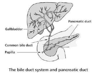
This is a medical examination performed to diagnose and treat disorders of the bile duct, gallbladder, pancreas and liver.
The most common reasons to do an ERCP are jaundice (yellowing of the skin or eyes) or abnormal liver tests or if a scan (ultrasound or CT scan) shows a blockage of the bile of pancreatic ducts. Blockages can be caused by stones, narrowing of the bile ducts and growths or cancers of the pancreas and bile ducts.
During an ERCP, stents (small plastic or metal tubes) can be inserted into the bile ducts to allow bile to drain into the intestine. If indicated a therapeutic procedure can then be undertaken. This may involve the removal of gallstones by cutting a larger opening in the bile duct (sphincterotomy) allowing the stones to fall out. An ERCP can give more information about the pancreas and bile ducts and brushings and biopsies (specimens of cells) can be taken from the bile ducts or the pancreas for analysis.
An ERCP usually lasts between 30 and 60 minutes depending on its complexity.
It would be helpful to bring a list of your current medications.
It is advisable to take your regular medications with a small amount of water no less than two hours before your test i.e. blood pressure tablets. Except:
Please make sure that you are told when to stop this medication by the doctor who referred you for this procedure. The level of the blood test (INR) must be <1.3 on the day of your procedure. If necessary your GP surgery can seek further specialist advice from the hospital haematology department.
Furthermore, you should avoid the following activities after the procedure:
Please check with your travel insurance if you wish to travel within 4 weeks of your procedure.
If you start to feel unwell or feverish, or develop severe abdominal pain, you must contact your GP or the local GP Emergency Service as soon as possible, it may be necessary to re-admit you to hospital.
You may resume a normal diet when able to do so.
After the procedure your throat may feel sore for up to twenty-four hours. The indication for this test will have been determined by previous tests, such as an ultrasound scan, and considered to be the safest way to help you. However, an ERCP is not risk free.
You should be aware of the following possible complications:
Bennet, J.R (1981). Therapeutic Endoscopy. Chapman and Hall.
Doctor Online (1999). ERCP www.doctoronline.nhs.uk
Gelton, P. and Williams, C (1997). Practical Gastrointestinal Endoscopy. Blackwell Science Ltd.
Hadley, A and Martin, D. Having an ERCP: A guide to the test.
Keymed.
“Having an ERCP” Guy’s and St Thomas’ NHS Foundation Trust 2018. Leaflet number: 2559/VER4 Accessed 24/07/2019.
© North Bristol NHS Trust. This edition published January 2024. Review due January 2027. NBT002079
If you are unable to attend your appointment please let us know as soon as possible. You can also contact the Imaging Department Contact Centre if you wish to change or discuss your appointment.
Telephone: 0117 414 8989
See the impact we make across our hospitals and how you can be a part of it.
Your doctor has requested that you have a rhizolysis procedure. We hope the following information will answer some of the questions you may have about this procedure.
This is a technique that deliberately damages the nerve that supplies the facet joint. These nerves are responsible for transmitting the pain that is caused by the facet joints.
If you have had success from facet joint injections in the past but the effects have not lasted, this procedure may offer improved long-term pain relief. The effect of this procedure may take longer to act (a few weeks), however could last up to a year and beyond.
The doctor in charge of your case feels this is an appropriate procedure for you. However you will have the opportunity for your opinion to be taken into account; if you do not want the procedure carried out then you can decide against it.
Finally we hope this information is helpful. If you have any questions either before, during or after the procedure the staff in the Imaging department will be happy to answer them.
The telephone number for the Imaging department can be found on the appointment letter.
Journal of spinal disorders and techniques. 2011 Apr;24 (2) 69-75
Spine. 2008 May 20; 33 (12): 1291-7
Southmead Hospital, Bristol, BS10 5NB
See your appointment letter for the number to phone with any queries you may have.
If you or the individual you are caring for need support reading this information please ask a member of staff for advice.
© North Bristol NHS Trust. This edition published January 2024. Review due January 2027. NBT002796
The Radiology Department may also be called the X-ray or Imaging Department. It is the facility in the hospital where radiological examinations are carried out using a variety of equipment.
Radiologists are the doctors who are specially trained to interpret the images and carry out more complex examinations. Radiologists are supported by Radiographers who are highly trained to carry out the examinations. You have been referred for an MRI scan. The results will go back to the clinic that referred you.
The MRI scanner is a circular tube which is open at both ends. MRI uses a very strong magnet to produce cross-sectional images of the body.
The magnet in the scanner collects information and sends it to the computer in the control room which displays your images.
The scanner is very noisy while it collects the images, and it is important you stay still so clear pictures can be taken.
MRI does not use any form of X-ray. MRI is now the alternative for some X-ray and operative procedures. The images produced by MRI are very detailed and show bones and soft tissue.
It may not be possible to have an MRI if you have a metal implanted device, this may include:
Please let the MRI unit know as soon as possible if you have any implanted devices. The phone number is on your appointment letter.
Before your MRI scan, you will be asked some questions to check that you are safe to enter the scan room. People with dental fillings, bridges, joint replacements or stents can all be scanned if you have had them for at least 6 weeks. The Radiographers will need to be aware of these so they can optimise your scan.
If you are pregnant at the time of your MRI scan, this should be discussed with the person referring you before attending the scan. If it is deemed necessary to still have your scan while you are pregnant, this will be discussed with the Radiologist and the appropriate safety precautions taken.
If you suffer from claustrophobia, we have experienced staff who can use a variety of techniques to help you through the scan. For severe claustrophobia, you may also be able to source sedatives from your GP however you will not be able to drive after taking these.
Most MRI examinations do not require you to have an injection but in some situations, it may be deemed beneficial by the consultant Radiologist.
The contrast is injected into a vein and can help to give more information which can contribute to your diagnosis.
If you are having an arthrogram the contrast will be injected into the joint space.
MRI contrast is not the same as X-ray contrast.
Children under 16 can be accompanied at all stages of the examination by an appropriate adult. Everyone coming into the scan room will be asked the safety questions about metal and implants, and be expected to change into appropriate clothing if required.
Please be aware that we are not able to supervise children while you have your scan.
You can be in the MRI department for between 20 minutes to 2 hours depending on your scan. Each area of the body takes approximately 20 minutes to scan.
Whilst we endeavour to ensure that you are seen at your appointment time sometimes emergencies may have to take priority. We ask for your patience and understanding should this occur, we will keep you informed of any delays.
You will not get any results at the time of your scan. A radiologist will report on your images and send the results to the doctor or clinic that requested the scan. You will receive the results from the clinician that requested your scan.
Royal College of Radiologists (RCR) 2001 Information for Adult Patients having an MRI Scan Available from: www.RCR. ac.uk [accessed April 2006].
Please do not hesitate to ask questions either before or after your scan. Contact details can be found on your appointment card.
© North Bristol NHS Trust. This edition published July 2024. Review due July 2027. NBT002009
See the impact we make across our hospitals and how you can be a part of it.